- June 11, 2025 - The NSD imagery data (one additional 7T fMRI scan session) are scheduled to be publicly released on September 1, 2025.
- March 11, 2025 - The NSD synthetic data (one additional 7T fMRI scan session) have now been publicly released.
- April 2, 2024: Take the NSD / large-scale neuroimaging dataset anonymous survey! Deadline May 15, 2024.
- January
16, 2023: Announcing that NSD data are used as part of the
2023 Algonauts
Challenge!
- January
13, 2023: A list of papers and pre-prints using NSD data
added below.
- December 16, 2021: The NSD data paper is now published.
- September
3, 2021: The NSD dataset
is now publicly available.
 For a formal description of
the dataset, please see the NSD data paper:
For a formal description of
the dataset, please see the NSD data paper:Allen, E.J., St-Yves, G., Wu, Y., Breedlove, J.L., Prince, J.S., Dowdle, L.T., Nau, M., Caron, B., Pestilli, F., Charest, I., Hutchinson, J.B., Naselaris, T.*, Kay, K.* A massive 7T fMRI dataset to bridge cognitive neuroscience and artificial intelligence. Nature Neuroscience (2021).
For a formal description of the NSD synthetic addition, please see the NSD synthetic paper:
Gifford, A.T., Cichy, R.M., Naselaris, T., Kay, K. A 7T fMRI dataset of synthetic images for out-of-distribution modeling of vision. arXiv (2025).In addition, here is a brief (20 min) talk presenting the NSD dataset (given to the MRC CBU Chaucer Club).
If you would like to access the NSD dataset, please fill out this short NSD Data Access Agreement. Upon completion, you will receive a link to the NSD Data Manual which provides technical information, a comprehensive description of the data files, and information on how to access the dataset.
The NSD dataset is a collaborative effort between PI Kendrick Kay and PI Thomas Naselaris, and the NSD data effort included contributions from the following researchers:
 Funding for the NSD dataset was graciously
provided by a grant
from the National Science Foundation.
Funding for the NSD dataset was graciously
provided by a grant
from the National Science Foundation.Papers and pre-prints
Here are links to papers that use NSD data.
- Fractional Ridge Regression: a Fast, Interpretable Reparameterization of Ridge Regression. Rokem, A. & Kay, K. GigaScience (2020).
- Extensive sampling for complete models of individual brains. Naselaris, T., Allen, E., & Kay, K. Current Opinion in Behavioral Sciences (2021).
- A massive 7T fMRI dataset to bridge cognitive neuroscience and artificial intelligence. Allen, St-Yves, Wu, Breedlove, Prince, Dowdle, Nau, Caron, Pestilli, Charest, Hutchinson, Naselaris*, & Kay*. Nature Neuroscience (2022).
- NeuroGen: activation optimized image synthesis for discovery neuroscience.Gu, Z., Jamison, K.W., Khosla, M., Allen, E.J., Wu, Y., Naselaris, T., Kay, K., Sabuncu, M.R., Kuceyeski, A.NeuroImage (2022).
- Non-Neural Factors Influencing BOLD Response Magnitudes within Individual Subjects.Kurzawski, J.W., Gulban, O.F., Jamison, K., Winawer, J.*, Kay, K.* Journal of Neuroscience (2022).
- Improving the accuracy of single-trial fMRI response estimates using GLMsingle.Prince, J.S., Charest, I., Kurzawski, J.W., Pyles, J.A., Tarr, M.J., Kay, K.N. eLife (2022).
- Personalized visual encoding model construction with small data.Zijin Gu, Keith Jamison, Mert Sabuncu, and Amy KuceyeskiCommunications Biology (2022).
- Selectivity for food in human ventral visual cortex.Nidhi Jain, Aria Wang, Margaret M. Henderson, Ruogu Lin, Jacob S. Prince, Michael J. Tarr, and Leila WehbeCommunications Biology (2023).
- Short-term plasticity in the human visual thalamus.Jan W Kurzawski, Claudia Lunghi, Laura Biagi, Michela Tosetti, Maria Concetta Morrone, Paola BindaeLife (2022).
- Color-biased regions in the ventral visual pathway are food selective.Pennock, I.M.L., Racey, C., Allen, E.J., Wu, Y., Naselaris, T., Kay, K.N., Franklin, A., Bosten, J.M.Current Biology (2022).
- Multiple Traces and Altered Signal-to-Noise in Systems Consolidation: Complementary Evidence from the 7T fMRI Natural Scenes Dataset.Vanasse, T.J., Boly, M., Allen, E.J., Wu, Y., Naselaris, T., Kay, K., Cirelli, C., Tononi, G.PNAS (2022).
- The risk of bias in data denoising methods: examples from neuroimaging.Kay, K.PLoS One (2022).
- A Highly Selective Response to Food in Human Visual Cortex Revealed by Hypothesis-Free Voxel Decomposition.Meenakshi Khosla, N. Apurva Ratan Murty, Nancy G KanwisherCurrent Biology (2022).
- See commentary:
Visual cortex: Big data analysis uncovers food specificity.Michael M. Bannert and Andreas BartelsCurrent Biology (2022). - Low-level tuning biases in higher visual cortex reflect the semantic informativeness of visual features.Margaret Henderson, Michael J. Tarr, Leila WehbeJournal of Vision (2023).
- Re-expression of CA1 and entorhinal activity patterns preserves temporal context memory at long timescales.Futing Zou, Wanjia Guo, Emily J. Allen, Yihan Wu, Ian Charest, Thomas Naselaris, Kendrick Kay, Brice A. Kuhl, J. Benjamin Hutchinson, Sarah DuBrowNature Communications (2023).
- A texture statistics encoding model reveals hierarchical feature selectivity across human visual cortex.Margaret M. Henderson, Michael J. Tarr, Leila WehbeJournal of Neuroscience (2023).
- Natural scene sampling reveals reliable coarse-scale orientation tuning in human V1.Roth, Z.N., Kay, K.*, Merriam, E.P.*Nature Communications (2022).
- Representations in human primary visual cortex drift over time.Roth, Z.N., Merriam, E.P.Nature Communications (2023).
- Human brain responses are modulated when exposed to optimized natural images or synthetically generated imagesZijin Gu, Keith Jamison, Mert R. Sabuncu, and Amy KuceyeskiCommunications Biology (2023).
- Brain-optimized deep neural networks of human visual areas learn non-hierarchical representations.St-Yves, G., Allen, E.J., Wu, Y., Kay, K.*, Naselaris, T.* Nature Communications (2023).
- Natural scene reconstruction from fMRI signals using generative latent diffusionFurkan Ozcelik and Rufin VanRullen.Scientific Reports (2023).
- Better models of human high-level visual cortex emerge from natural language supervision with a large and diverse dataset.Wang, A.Y., Kay, K., Naselaris, T., Tarr, M.J., Wehbe, L.Nature Machine Intelligence (2023).
- Encoding of Visual Objects in the Human Medial Temporal LobeYue Wang, Runnan Cao and Shuo WangJournal of Neuroscience (2024).
- Mind-bridge: reconstructing visual images based on diffusion model from human brain activityQing Liu, Hongqing Zhu, Ning Chen, Bingcang Huang, Weiping Lu & Ying WangSignal, Image and Video Processing (2024)
- A unifying framework for functional organization in early and higher ventral visual cortexEshed Margalit, Hyodong Lee, Dawn Finzi, James J. DiCarlo, Kalanit Grill-Spector, Daniel L.K. YaminsNeuron (2024).
- Natural scenes reveal diverse representations of 2D and 3D body pose in the human brainHongru Zhu, Yijun Ge, Alexander Bratch, Alan Yuille, Kendrick Kay, Daniel KerstenPNAS (2024).
- Retrieving and reconstructing conceptually similar images from fMRI with latent diffusion models and a neuro-inspired brain decoding modelMatteo Ferrante, Tommaso Boccato, Luca Passamonti, and Nicola ToschiJournal of Neural Engineering (2024).
- Large-scale parameters framework with large convolutional kernel for encoding visual fMRI activity informationShuxiao Ma, Linyuan Wang, Senbao Hou, Chi Zhang, Bin YanCerebral Cortex (2024).
- Primate brain: A unique connection between dorsal and ventral visual cortexJason D. YeatmanCurrent Biology (2024).
- Frontostriatal salience network expansion in individuals in depressionCharles J. Lynch, Immanuel G. Elbau, Tommy Ng, Aliza Ayaz, Shasha Zhu, Danielle Wolk, Nicola Manfredi, Megan Johnson, Megan Chang, Jolin Chou, Indira Summerville, Claire Ho, Maximilian Lueckel, Hussain Bukhari, Derrick Buchanan, Lindsay W. Victoria, Nili Solomonov, Eric Goldwaser, Stefano Moia, Cesar Caballero-Gaudes, Jonathan Downar, Fidel Vila-Rodriguez, Zafiris J. Daskalakis, Daniel M. Blumberger, Kendrick Kay, Amy Aloysi, Evan M. Gordon, Mahendra T. Bhati, Nolan Williams, Jonathan D. Power, Benjamin Zebley, Logan Grosenick, Faith M. Gunning & Conor ListonNature (2024).
- Contrastive learning explains the emergence and function of visual category-selective regionsJacob S. Prince, George A. Alvarez, and Talia KonkleScience Advances (2024).
- A large-scale examination of inductive biases shaping high-level visual representation in brains and machinesColin Conwell, Jacob S. Prince, Kendrick N. Kay, George A. Alvarez & Talia KonkleNature Communications (2024).
- NeuralDiffuser: Neuroscience-inspired Diffusion Guidance for fMRI Visual ReconstructionHaoyu Li, Hao Wu, Badong ChenIEEE Transactions on Image Processing (2025).
- Unraveling the Differential Efficiency of Dorsal and Ventral Pathways in Visual Semantic DecodingWei Huang, Ying Tang, Sizhuo Wang, Jingpeng Li, Kaiwen Cheng, and Hongmei YanInternational Journal of Neural Systems (2025).
- The human social cognitive network contains multiple regions within the amygdala. Edmonds, D., Salvo, J.J., Anderson, N., Lakshman, M., Yang, Q., Kay, K., Zelano, C., Braga, R.M. Science Advances (2024).
- Situating the salience and parietal memory networks in the context of multiple parallel distributed networks using precision functional mapping. Kwon, Y.H., Salvo, J.J., Anderson, N.L., Edmonds, D., Holubecki, A.M., Lakshman, M., Yoo, K., Yeo B.T.T., Kay, K., Gratton, C., Braga, R.M. Cell Reports (2025).
- NeuroFusionNet: cross-modal modeling from brain activity to visual understanding. Kehan Lang, Jianwei Fang, Guangyao Su. Frontiers in Computational Neuroscience (2025).
- Memory recall: Retrieval-Augmented mind reconstruction for brain decoding. Yuxiao Zhao, Guohua Dong, Lei Zhu, Xiaomin Ying. Information Fusion (2025).
Here are links to conference papers and pre-prints that use NSD data.
- What can 5.17 billion regression fits tell us about artificial models of the human visual system?Colin Conwell, Jacob S. Prince, George A. Alvarez, Talia KonkleNeurIPS SVRHM workshop (2021).
- Large-Scale Benchmarking of Diverse Artificial Vision Models in Prediction of 7T Human Neuroimaging Data.Colin Conwell, Jacob S. Prince, George A. Alvarez, Talia KonklebioRxiv (2022).
- High-level visual areas act like domain-general filters with strong selectivity and functional specialization.Meenakshi Khosla, Leila WehbebioRxiv (2022).
- Semantic scene descriptions as an objective of human visionDoerig, A., Kietzmann, T.C., Allen, E., Wu, Y., Naselaris, T., Kay, K., Charest, I.arXiv (2022).
- Mind Reader: Reconstructing complex images from brain activities.Sikun Lin, Thomas Sprague, Ambuj K Singh.arXiv (2022).
- High-resolution image reconstruction with latent diffusion models from human brain activity.Takagi, Y., Nishimoto, S.bioRxiv (2022).
- Decoding natural image stimuli from fMRI data with a surface-based convolutional network.Zijin Gu, Keith Jamison, Amy Kuceyeski, Mert SabuncuarXiv (2022).
- Sample Reweighting for Label Denoising of Neural Activity DataDongfang Xu, Rong ChenIEEE/EMBS Conference on Neural Engineering (2023)
- The Algonauts Project 2023 Challenge: How the Human Brain Makes Sense of Natural Scenes.A.T. Gifford, B. Lahner, S. Saba-Sadiya, M.G. Vilas, A. Lascelles, A. Oliva, K. Kay, G. Roig, R.M. Cichy.arXiv (2023).
- Neural Selectivity for Real-World Object Size In Natural ImagesAndrew F. Luo, Leila Wehbe, Michael J. Tarr, Margaret M. HendersonbioRxiv (2023)
- MindDiffuser: Controlled Image Reconstruction from Human Brain Activity with Semantic and Structural DiffusionYizhuo Lu, Changde Du, Dianpeng Wang, Huiguang HearXiv (2023).
- The transition from vision to language: distinct patterns of functional connectivity for sub-regions of the visual word form areaMaya Yablonski, Iliana I Karipidis, Emily Kubota, Jason D YeatmanbioRxiv (2023).
- Reconstructing seen images from human brain activity via guided stochastic searchReese Kneeland, Jordyn Ojeda, Ghislain St-Yves, Thomas NaselarisarXiv (2023).
- BrainCLIP: Bridging Brain and Visual-Linguistic Representation Via CLIP for Generic Natural Visual Stimulus DecodingYulong Liu, Yongqiang Ma, Wei Zhou, Guibo Zhu, Nanning ZhengarXiv (2023).
- Brain Captioning: Decoding human brain activity into images and textMatteo Ferrante, Furkan Ozcelik, Tommaso Boccato, Rufin VanRullen, Nicola ToschiarXiv (2023).
- A Unifying Principle for the Functional Organization of Visual CortexEshed Margalit, Hyodong Lee, Dawn Finzi, James J. DiCarlo, Kalanit Grill-Spector, Daniel L. K. YaminsarXiv (2023).
- Reconstructing the Mind’s Eye: fMRI-to-Image with Contrastive Learning and Diffusion PriorsPaul S. Scotti*, Atmadeep Banerjee*, Jimmie Goode, Stepan Shabalin, Alex Nguyen, Ethan Cohen, Aidan J. Dempster, Nathalie Verlinde, Elad Yundler, David Weisberg, Kenneth A. Norman*, and Tanishq Mathew Abraham*arXiv (2023).
- Brain Dissection: fMRI-trained Networks Reveal Spatial Selectivity in the Processing of Natural ImagesGabriel H. Sarch, Michael J. Tarr, Katerina Fragkiadaki, Leila WehbearXiv (2023).
- Second Sight: Using brain-optimized encoding models to align image distributions with human brain activityReese Kneeland, Jordyn Ojeda, Ghislain St-Yves, Thomas NaselarisarXiv (2023).
- Brain Diffusion for Visual Exploration: Cortical Discovery using Large Scale Generative Models.Andrew F. Luo, Margaret M. Henderson, Leila Wehbe, Michael J. TarrarXiv (2023). [NeurIPS 2023 (Oral)].
- Improving visual image reconstruction from human brain activity using latent diffusion models via multiple decoded inputs.Yu Takagi, Shinji NishimotoarXiv (2023).
- DreamCatcher: Revealing the Language of the Brain with fMRI using GPT EmbeddingSubhrasankar Chatterjee, Debasis SamantaarXiv (2023).
- What can 1.8 billion regressions tell us about the pressures shaping high-level visual representation in brains and machines?Colin Conwell, Jacob S. Prince, Kendrick N. Kay, George A. Alvarez, Talia KonklebioRxiv (2023)
- THE ALGONAUTS PROJECT 2023 CHALLENGE: UARK-UALBANY TEAM SOLUTIONXuan Bac Nguyen, Xudong Liu, Xin Li, Khoa LuuarXiv (2023)
- Memory Encoding ModelHuzheng Yang, James Gee, Jianbo ShiarXiv (2023)
- Applicability of scaling laws to vision encoding modelsTakuya Matsuyama, Kota S Sasaki, Shinji NishimotoarXiv (2023).
- A contrastive coding account of category selectivity in the ventral visual streamJacob S. Prince, George A. Alvarez, Talia KonklebioRxiv (2023).
- Predicting brain activity using TransformersHossein Adeli, Sun Minni, Nikolaus KriegeskortebioRxiv (2023).
- A Parameter-efficient Multi-subject Model for Predicting fMRI ActivityConnor Lane, Gregory KiararXiv (2023).
- Expansion of a frontostriatal salience network in individuals with depressionCharles J. Lynch, I. Elbau, Tommy Ng, Aliza Ayaz, Shasha Zhu, Nicola Manfredi, Megan A. Johnson, Daniel L Wolk, Jonathan D. Power, E. Gordon, Kendrick Norris Kay, A. Aloysi, Stefano Moia, C. Caballero-Gaudes, L. Victoria, N. Solomonov, E. Goldwaser, Benjamin Zebley, L. Grosenick, J. Downar, F. Vila-Rodriguez, Z. Daskalakis, D. Blumberger, N. Williams, F. Gunning, C. ListonbioRxiv (2023).
- UniBrain: Unify Image Reconstruction and Captioning All in One Diffusion Model from Human Brain ActivityWeijian Mai, Zhijun ZhangarXiv (2023).
- A Multimodal Visual Encoding Model Aided by Introducing Verbal Semantic InformationMa Shuxiao, Wang Linyuan, Yan BinarXiv (2023).
- Through their eyes: multi-subject Brain Decoding with simple alignment techniquesMatteo Ferrante, Tommaso Boccato, and Nicola ToschiarXiv (2023).
- Direct perception of affective valence from visionSaeedeh Sadeghi, Zijin Gu, Eve DeRosa, Amy Kuceyeski, Adam K. AndersonpsyArXiv (2023).
- Dissociable contributions of the medial parietal cortex to recognition memorySeth R. Koslov, Joseph W. Kable, & Brett L. FosterbioRxiv (2023).
- UNIDIRECTIONAL BRAIN-COMPUTER INTERFACE: ARTIFICIAL NEURAL NETWORK ENCODING NATURAL IMAGES TO fMRI RESPONSE IN THE VISUAL CORTEXRuixing Liang, Xiangyu Zhang, Qiong Li, Lai Wei, Hexin Liu, Avisha Kumar, Kelley M. Kempski Leadingham, Joshua Punnoose, Leibny Paola Garcia, Amir ManbachiarXiv (2023).
- Cortical and subcortical brain networks predict prevailing heart rateAmy Isabella Sentis, Javier Rasero, Peter J. Gianaros, Timothy D. VerstynenbioRxiv (2023).
- DREAM: Visual Decoding from REversing HumAn Visual SysteMWeihao Xia, Raoul de Charette, Cengiz Oztireli, Jing-Hao XuearXiv (2023).
- BrainSCUBA: Fine-Grained Natural Language Captions of Visual Cortex SelectivityAndrew F. Luo, Margaret M. Henderson, Michael J. Tarr, Leila WehbearXiv (2023).
- IDENTIFYING INTERPRETABLE VISUAL FEATURES INARTIFICIAL AND BIOLOGICAL NEURAL SYSTEMSDavid Klindt, Sophia Sanborn, Francisco Acosta, Fr ́ed ́eric Poitevin, Nina MiolanearXiv (2023).
- fMRI-PTE: A Large-scale fMRI Pretrained Transformer Encoder for Multi-Subject Brain Activity DecodingXuelin Qian, Yun Wang, Jingyang Huo, Jianfeng Feng, Yanwei FuarXiv (2023).
- BRAIN DECODING: TOWARD REAL-TIME RECONSTRUCTION OF VISUAL PERCEPTIONYohann Benchetrit, Hubert Banville, Jean-Remi KingarXiv (2023).
- Soft Matching Distance: A metric on neural representations that captures single-neuron tuningMeenakshi Khosla, Alex H. WilliamsarXiv (2023).
- Brainformer: Modeling MRI Brain Functions to Machine VisionXuan-Bac Nguyen , Xin Li, Samee U. Khan, Khoa LuuarXiv (2023)
- Brain Decodes Deep NetsHuzheng Yang, James Gee*, Jianbo Shi*arXiv (2023)
- OneLLM: One Framework to Align All Modalities with LanguageJiaming Han, Kaixiong Gong, Yiyuan Zhang, Jiaqi Wang, Kaipeng Zhang,Dahua Lin, Yu Qiao, Peng Gao, Xiangyu YuearXiv (2023).
- Lite-Mind: Towards Efficient and Versatile Brain Representation NetworkZixuan Gong, Qi Zhang, Duoqian Miao, Guangyin Bao, Liang HuarXiv (2023).
- Multimodal decoding of human brain activity into images and textMatteo Ferrante, Tommaso Boccato, Furkan Ozcelik, Rufin VanRullen, Nicola ToschiNeurIPS (2023).
- ALIGNING BRAIN FUNCTIONS BOOSTS THE DECODING OF VISUAL SEMANTICS IN NOVEL SUBJECTSAlexis Thual, Yohann Benchetrit, Felix Geilert, Jeremy Rapin, Iurii Makarov, Hubert Banville, Jean-Remi KingarXiv (2023).
- Brain-optimized inference improves reconstructions of fMRI brain activityReese Kneeland, Jordyn Ojeda, Ghislain St-Yves, Thomas NaselarisarXiv (2023).
- Body Cosmos: An Immersive Experience Driven by Real-Time Bio-DataRem RunGu Lin; Yongen Ke; Kang ZhangIEEE VIS Arts Program (VISAP) (2023).
- MinD-3D: Reconstruct High-quality 3D objects in Human BrainJianxiong Gao, Yuqian Fu, Yun Wang, Xuelin Qian, Jianfeng Feng, Yanwei FuarXiv (2023).
- A single computational objective drives specialization of streams in visual cortexDawn Finzi, Eshed Margalit, Kendrick Kay, Daniel L. K. Yamins, Kalanit Grill-SpectorbioRxiv (2023).
- Evaluation of Representational Similarity Scores Across Human Visual CortexFrancisco Acosta, Colin Conwell, Sophia Sanborn, David A. Klindt, Nina MiolaneNeurIPS (2023).
- A randomized algorithm to solve reduced rank operator regressionGiacomo Turri*, Vladimir Kostic*, Pietro Novelli*, and Massimiliano PontilarXiv (2023).
- Aligned with LLM: a new multi-modal training paradigm for encoding fMRI activity in visual cortexShuxiao Ma, Linyuan Wang, Senbao Hou, Bin YanarXiv (2024).
- Recent statistics shift object representations in parahippocampal cortexSolomon, S.H., Kay, K., & Schapiro, A.C.bioRxiv (2024).
- Parsing Brain Network Specialization: A Replication and Expansion of Wang et al. (2014)Madeline Peterson, Dorothea L. Floris, and Jared A. NielsenbioRxiv (2024).
- CLIP-MUSED: CLIP-GUIDED MULTI-SUBJECT VISUAL NEURAL INFORMATION SEMANTIC DECODINGQiongyi Zhou, Changde Du, Shengpei Wang and Huiguang HeICLR (2024).
- NeuralDiffuser: Controllable fMRI Reconstruction with Primary Visual Feature Guided DiffusionHaoyu Li, Hao Wu, Badong ChenarXiv (2024).
- Visual Image Reconstruction from Human Brain Activity using Linear Image Decoders plus Nonlinear Noise SuppressionQiang LibioRxiv (2024).
- See Through Their Minds: Learning Transferable Neural Representation from Cross-Subject fMRI Yulong Liu, Yongqiang Ma, Guibo Zhu, Haodong Jing, and Nanning ZhengarXiv (2024).
- Inter-individual and inter-site neural code conversion and image reconstruction without shared stimuliHaibao Wang, Jun Kai Ho, Fan L. Cheng, Shuntaro C. Aoki, Yusuke Muraki, Misato Tanaka, Yukiyasu KamitaniarXiv (2024).
- NeuroPictor: Refining fMRI-to-Image Reconstruction via Multi-individual Pretraining and Multi-level ModulationJingyang Huo, Yikai Wang, Xuelin Qian, Yun Wang, Chong Li, Jianfeng Feng, Yanwei FuarXiv (2024).
- Psychometry: An Omnifit Model for Image Reconstruction from Human Brain ActivityRuijie Quan, Wenguan Wang, Zhibo Tian, Fan Ma, Yi YangarXiv (2024).
- Reconstructing Retinal Visual Images from 3T fMRI Data Enhanced by Unsupervised LearningYujian Xiong, Wenhui Zhu, Zhong-Lin Lu, Yalin WangarXiv (2024).
- Unified Multimodal Decoding of Brain SignalsWeihao Xia, Raoul de Charette, Cengiz Oztireli, Jing-Hao XuearXiv (2024).
- MindBridge: A Cross-Subject Brain Decoding FrameworkShizun Wang, Songhua Liu, Zhenxiong Tan, Xinchao WangarXiv (2024).
- Functional Brain-to-Brain Transformation with No Shared DataNavve Wasserman, Roman Beliy, Roy Urbach, and Michal IraniarXiv (2024).
- MindTuner: Cross-Subject Visual Decoding with Visual Fingerprint and Semantic CorrectionZixuan Gong, Qi Zhang, Guangyin Bao, Lei Zhu, Ke Liu, Liang Hu, Duoqian MiaoarXiv (2024).
- Wills Aligner: A Robust Multi-Subject Brain Representation LearnerGuangyin Bao, Zixuan Gong, Qi Zhang, Jialei Zhou, Wei Fan, Kun Yi, Usman Naseem, Liang Hu, Duoqian MiaoarXiv (2024).
- Integrated Gradient Correlation: a Dataset-wise Attribution MethodPierre Lelièvre, Chien-Chung ChenarXiv (2024).
- Disentangling signal and noise in neural responses through generative modelingKay, K., Prince, J.S., Gebhart, T., Tuckute, G., Zhou, J., Naselaris, T., Schutt, H.bioRxiv (2024).
- Neuro-Vision to Language: Image Reconstruction and Language enabled Interaction via Brain RecordingsGuobin Shen, Dongcheng Zhao, Xiang He, Linghao Feng, Yiting Dong, Jihang Wang, Qian Zhang and Yi ZengarXiv (2024).
- Automating the Diagnosis of Human Vision Disorders by Cross-modal 3D GenerationLi Zhang, Yuankun Yang, Ziyang Xie, Zhiyuan Yuan, Jianfeng Feng, Xiatian Zhu, and Yu-Gang JiangarXiv (2024)
- MindShot: Brain Decoding Framework Using Only One ImageShuai Jiang, Zhu Meng, Delong Liu, Haiwen Li, Fei Su and Zhicheng ZhaoarXiv (2024).
- MindFormer: A Transformer Architecture for Multi-Subject Brain Decoding via fMRIInhwa Han, Jaayeon Lee, Jong Chul YearXiv (2024).
- MindSemantix: Deciphering Brain Visual Experiences with a Brain-Language ModelZiqi Ren, Jie Li, Xuetong Xue, Xin Li, Fan Yang, Zhicheng Jiao, Xinbo GaoarXiv (2024).
- The Wisdom of a Crowd of Brains: A Universal Brain EncoderRoman Beliy, Navve Wasserman, Amit Zalcher, Michal IraniarXiv (2024)
- Neuro-Vis: Guided Complex Image Reconstruction from Brain Signals Using Multiple Semantic and Perceptual ControlsGabriela M. Balisacan, Anne Therese A. PauloCMLDS '24: Proceedings of the International Conference on Computing, Machine Learning and Data Science (2024)
- Privileged representational axes in biological and artificial neural networksMeenakshi Khosla, Alex H Williams, Josh McDermott, Nancy KanwisherbioRxiv (2024).
- BrainMAE: A Region-aware Self-supervised Learning Framework for Brain SignalsYifan Yang, Yutong Mao, Xufu Liu, Xiao LiuarXiv (2024).
- AlignedCut: Visual Concepts Discovery on Brain-Guided Universal Feature SpaceHuzheng Yang, James Gee*, Jianbo Shi*arXiv (2024)
- MindLDM: Reconstruct Visual Stimuli from fMRI Using Latent Diffusion ModelJunhao Guo; Chanlin Yi; Fali Li; Peng Xu; Yin Tian2024 IEEE International Conference on Computational Intelligence and Virtual Environments for Measurement Systems and Applications (CIVEMSA) (2024)
- NeuroBind: Towards Unified Multimodal Representations for Neural SignalsFengyu Yang, Chao Feng, Daniel Wang, Tianye Wang, Ziyao Zeng, Zhiyang Xu, Hyoungseob Park, Pengliang Ji, Hanbin Zhao, Yuanning Li, Alex WongarXiv (2024).
- Neural 3D decoding for human vision diagnosisLi Zhang, Yuankun Yang, Ziyang Xie, Zhiyuan Yuan, Jianfeng Feng, Xiatian Zhu, Yu-Gang JiangarXiv (2024).
- Temporal asymmetry of neural representations predicts memory decisionsZhifang Ye, Yufei Zhao, Emily J. Allen, Thomas Naselaris, Kendrick Kay, J. Benjamin Hutchinson, Brice A. KuhlbioRxiv (2024).
- Hierarchical Quantum Control Gates for Functional MRI UnderstandingXuan-Bac Nguyen, Hoang-Quan Nguyen, Hugh Churchill, Samee U. Khan, Khoa LuuarXiv (2024).
- UNIVERSAL DIMENSIONS OF VISUAL REPRESENTATIONZirui Chen & Michael F. BonnerarXiv (2024).
- FedMinds: Privacy-Preserving Personalized Brain Visual DecodingGuangyin Bao, Duoqian MiaoarXiv (2024).
- Efficient fMRI and Textual Alignment for ImageReconstruction from Human Brain ActivityBich-Nga Pham, Trong-Tai Dam Vu, Anh-Khoa Nguyen Vu, Vinh-Tiep NguyenResearch Square (2024).
- UNIVERSAL SCALE-FREE REPRESENTATIONS IN HUMAN VISUAL CORTEXRaj Magesh Gauthaman, Brice Ménard, Michael F. BonnerarXiv (2024).
- fMRI-3D: A Comprehensive Dataset for Enhancing fMRI-based 3D ReconstructionJianxiong Gao, Yuqian Fu, Yun Wang, Xuelin Qian, Jianfeng Feng, Yanwei FuarXiv (2024).
- Unsupervised alignment reveals structural commonalities and differences in neural representations of natural scenes across individuals and brain areasKen Takeda, Kota Abe, Jun Kitazono, Masafumi OizumibioRxiv (2024).
- Positive and Negative Retinotopic Codes in the Human HippocampusPeter A Angeli, Adam Steel, Edward H Silson, Caroline E RobertsonbioRxiv (2024).
- Decoding the Echoes of Vision from fMRI: Memory Disentangling for Past Semantic InformationRunze Xia, Congchi Yin, Piji LiarXiv (2024).
- On the Stability of Reduced-Rank Ridge RegressionDavid Degras, Thomas Chapalain, Bertrand ThirionEUSIPCO 2024 - 32nd European signal processing conference (2024).
- Generalizability analysis of deep learning predictions of human brain responses to augmented and semantically novel visual stimuliValentyn Piskovskyi, Riccardo Chimisso, Sabrina Patania, Tom Foulsham, Giuseppe Vizzari, and Dimitri OgnibenearXiv (2024).
- Contrastive Learning to Fine-Tune Feature Extraction Models for the Visual CortexAlex Mulrooney, Austin J. BrockmeierarXiv (2024).
- BRAIN MAPPING WITH DENSE FEATURES: GROUNDING CORTICAL SEMANTIC SELECTIVITY IN NATURAL IMAGES WITH VISION TRANSFORMERSAndrew F. Luo, Jacob Yeung, Rushikesh Zawar, Shaurya Dewan, Margaret M. Henderson, Leila Wehbe*, Michael J. Tarr*arXiv (2024).
- Retinotopic coding organizes the opponent dynamic between internally and externally oriented brain networksAdam Steel, Peter A. Angeli, Edward H. Silson, Caroline E. RobertsonbioRxiv (2024).
- Vicarious Somatotopic Maps Tile Visual CortexNicholas Hedger, Thomas Naselaris, Kendrick Kay, Tomas Knapen bioRxiv (2024).
- LINBRIDGE: A LEARNABLE FRAMEWORK FOR INTERPRETING NONLINEAR NEURAL ENCODING MODELSXiaohui Gao*, Yue Cheng*, Peiyang Li, Yijie Niu, Yifan Ren, Yiheng Liu, Haiyang Sun, Zhuoyi Li, Weiwei Xing, and Xintao HuarXiv (2024).
- Finding Shared Decodable Concepts and their Negations in the BrainCory Efird, Alex Murphy, Joel Zylberberg, Alona FyshearXiv (2024).
- Towards Neural Foundation Models for Vision: Aligning EEG, MEG, and fMRI Representations for Decoding, Encoding, and Modality ConversionMatteo Ferrante, Tommaso Boccato, Grigorii Rashkov, Nicola ToschiarXiv (2024).
- In silico discovery of representational relationships across visual cortexAlessandro T. Gifford, Maya A. Jastrzębowska, Johannes J.D. Singer, Radoslaw M. CichyarXiv (2024).
- Quantum-Brain: Quantum-Inspired Neural Network Approach to Vision-Brain UnderstandingHoang-Quan Nguyen, Xuan-Bac Nguyen, Hugh Churchill, Arabinda Kumar Choudhary, Pawan Sinha, Samee U. Khan, Khoa LuuarXiv (2024).
- OPTIMIZED TWO-STAGE AI-BASED NEURAL DECODING FOR ENHANCED VISUAL STIMULUS RECONSTRUCTION FROM FMRI DATALorenzo Veronese, Andrea Moglia, Luca Mainardi, Pietro CerveriarXiv (2024).
- UniBrain: A Unified Model for Cross-Subject Brain DecodingZicheng Wang, Zhen Zhao, Luping Zhou, Parashkev NachevarXiv (2024).
- Making Your Dreams A Reality: Decoding the Dreams into a Coherent Video Story from fMRI SignalsYanwei Fu, Jianxiong Gao, Baofeng Yang, Jianfeng FengarXiv (2025).
- BOLDreams: Dreaming with pruned in-silico fMRI Encoding Models of the Visual CortexUzair Hussain, Kamil UludagarXiv (2025).
- BRAINGUARD: Privacy-Preserving Multisubject Image Reconstructions from Brain ActivitiesZhibo Tian, Ruijie Quan, Fan Ma, Kun Zhan, Yi YangarXiv (2025).
- TopoNets: High performing vision and language models with brain-like topographyMayukh Deb, Mainak Deb, N. Apurva Ratan MurtyarXiv (2025).
- Scaling laws for decoding images from brain activityHubert Banville, Yohann Benchetrit, Stéphane d'Ascoli, Jérémy Rapin, Jean-Rémi KingarXiv (2025).
- TROI: CROSS-SUBJECT PRETRAINING WITH SPARSE VOXEL SELECTION FOR ENHANCED FMRI VISUAL DECODING. Ziyu Wang, Tengyu Pan, Zhenyu Li, Jianyong Wang, Xiuxing Li, and Ji Wu. arXiv (2025).
- LaVCa: LLM-assisted Visual Cortex Captioning. Takuya Matsuyama, Shinji Nishimoto, Yu Takagi. arXiv (2025).
- Braintransformer: Subject-Wise Patch Embed Transformer for Cross-Subject Brain Visual Information Decoding. Du Zongxin, Li Lin. 2024 21st International Computer Conference on Wavelet Active Media Technology and Information Processing (ICCWAMTIP)
- Talking to the brain: Using Large Language Models as Proxies to Model Brain Semantic Representation. Xin Liu, Ziyue Zhang, Jingxin Nie. arXiv (2025).
- MindLLM: A Subject-Agnostic and Versatile Model for fMRI-to-Text Decoding. Weikang Qiu, Zheng Huang, Haoyu Hu, Aosong Feng, Yujun Yan, Rex Ying. arXiv (2025).
- BrainChat: Interactive Semantic Information Decoding from fMRI Using Large-Scale Vision-Language Pretrained Models. Wanqiu Huang, Ke Ma, Tingyu Xie, Hongwei Wang. ICASSP 2025 - 2025 IEEE International Conference on Acoustics, Speech and Signal Processing (ICASSP) (2025).
- A 7T fMRI dataset of synthetic images for out-of-distribution modeling of vision. Alessandro T. Gifford, Radoslaw M. Cichy, Thomas Naselaris, Kendrick Kay. arXiv (2025).
- SEED: Towards More Accurate Semantic Evaluation for Visual Brain Decoding. Juhyeon Park, Peter Yongho Kim, Jiook Cha, Shinjae Yoo, and Taesup Moon. arXiv (2025).
- Monkey See, Model Knew: LARGE LANGUAGE MODELS ACCURATELY PREDICT VISUAL BRAIN RESPONSES IN HUMANS and NON-HUMAN PRIMATES. Colin Conwell, Emalie MacMahon, Akshay Jagadeesh, Kasper Vinken, Saloni Sharma, Jacob S. Prince, George A. Alvarez, Talia Konkle, Margaret Livingstone, and Leyla Isik. bioRxiv (2025).
- Optimal Transport for Brain-Image Alignment: Unveiling Redundancy and Synergy in Neural Information Processing. Yang Xiao, Wang Lu, Jie Ji, Ruimeng Ye, Gen Li, Xiaolong Ma, Bo Hui. arXiv (2025).
- Hierarchical Processing of Natural Scenes in the Human Pulvinar. Daniel R. Guest, Emily J. Allen, Kendrick N. Kay*, Michael J. Arcaro*. bioRxiv (2025).
- Enhancing 3T BOLD fMRI SNR using Unpaired 7T Data with Schrödinger Bridge Diffusion. Yujian Xiong, Xuanzhao Dong, Sebastian Waz, Wenhui Zhu, Negar Mallak, Zhong-Lin Lu, and Yalin Wang. arXiv (2025).
- From Eye to Mind: brain2text Decoding Reveals the Neural Mechanisms of Visual Semantic Processing. Feihan Feng & Jingxin Nie. arXiv (2025).
- CLIP-MSM: A Multi-Semantic Mapping Brain Representation for Human High-Level Visual Cortex. Guoyuan Yang, Mufan Xue, Ziming Mao, Haofang Zheng, Jia Xu, Dabin Sheng, Ruotian Sun, Ruoqi Yang, Xuesong Li. Proceedings of the AAAI Conference on Artificial Intelligence (2025).
- Probability Distribution Alignment and Low-Rank Weight Decomposition for Source-Free Domain Adaptive Brain Decoding. Ganxi Xu, Jinyi Long, Jia Zhang. arXiv (2025).
- Neural correlates of image memorability: Combining large-scale 7T fMRI with machine learning-based predictions. Mäki-Marttunen, V, Hagen, T., Espeseth, T. bioRxiv (2025).
- Predictive Coding Dynamics Enhance Model-Brain Similarity. Manshan Guo, Michael Samjatin, Bhavin Choksi, Sari Saba-Sadiya, Radoslaw M. Cichy, and Gemma Roig. ESANN 2025 proceedings, European Symposium on Artificial Neural Networks (2025).
- Multi-Task Vision Experts for Brain Captioning. Weihao Xia, Cengiz Oztirel. CVPR (2025).
- FreqSelect: Frequency-Aware fMRI-to-Image Reconstruction. Junliang Ye, Lei Wang, Md Zakir Hossain. arXiv (2025).
- Dynadiff: Single-stage Decoding of Images from Continuously Evolving fMRI. Marlène Careil*, Yohann Benchetrit*, Jean-Rémi King. arXiv (2025).
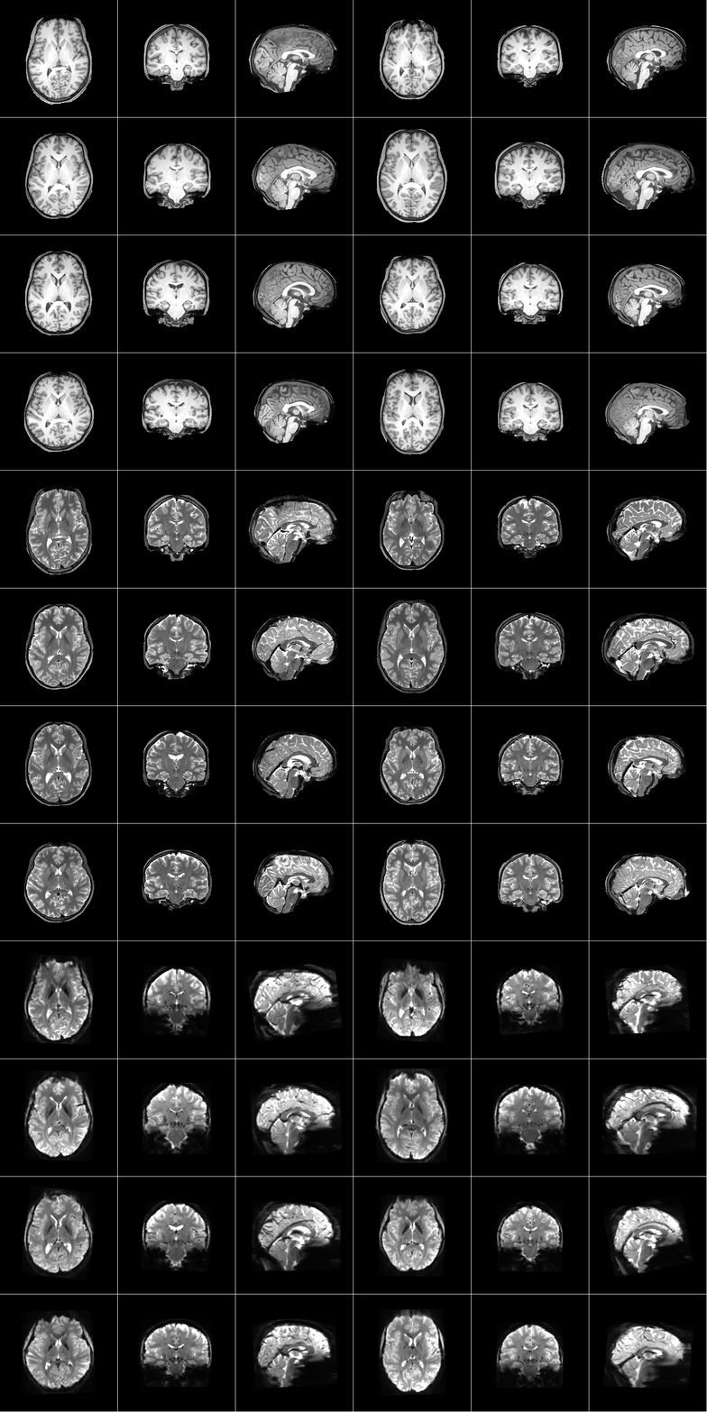
Sample data acquired on the 8 subjects:
T1-weighted 0.8-mm isotropic MRI,
T2-weighted 0.8-mm isotropic MRI,
T2*-weighted (EPI) 1.8-mm isotropic fMRI
